Integration of Computed Tomography (CT) Scans in Bone Stress Analysis
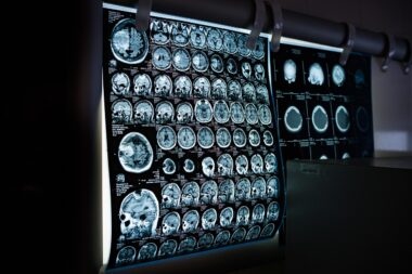
Biomechanics is a critical field of study that analyzes the mechanics of body systems. Understanding how internal and external forces impact bones can significantly affect healthcare. Among various imaging techniques, computed tomography (CT) scans play a vital role in bone stress analysis. These scans provide detailed cross-sectional images of bones, which are crucial for identifying stress fractures and other injuries. CT imaging allows for precise measurement of bone density, helping professionals assess the structural integrity of bones under various stress conditions. Integration of CT scans into biomechanical studies leads to improved diagnostic capabilities and enhanced treatment plans for patients. For example, those with osteoporosis can benefit from knowledge about their bone structure, reducing the risk of fractures. The comprehensive 3D models derived from CT scans let researchers visualize complex bone geometries. Consequently, this integration has profound implications, improving surgical planning and monitoring therapeutic outcomes. As technology advances, the capabilities of CT imaging continue to enhance the practice of biomechanics, revealing new avenues for research and development in clinical applications. Overall, understanding these techniques can revolutionize how we approach bone health and rehabilitation.
Advantages of Using CT in Biomechanics
One of the main advantages of using computed tomography is its ability to provide high-resolution images. Unlike traditional X-rays, CT scans allow for a more in-depth examination of the bone structures. This capability is beneficial in identifying fine details that conventional imaging might miss. Clinicians can visualize internal bone architecture and detect subtle changes that occur due to stress or injury. Additionally, the use of CT scans can facilitate more accurate modeling and simulation in biomechanics. By having precise measurements, researchers can simulate the mechanical behavior of bone tissues under various loads. This results in a deeper understanding of biomechanical properties and how they relate to specific injuries. Moreover, CT imaging allows for non-invasive analysis of bone stresses during a range of physical activities. This information is invaluable for sports medicine professionals who aim to optimize athletic performance while minimizing injury risks. It provides insights into how athletes’ bones respond to particular training regimens. Ultimately, leveraging CT imaging techniques enhances both preventive care and rehabilitative strategies, leading to improved outcomes for individuals across various demographics.
Computed tomography not only plays a significant role in detecting injuries but also transforms how rehabilitation strategies are developed. After significant injuries, such as fractures, a detailed understanding of the healing process is crucial. CT scans allow healthcare professionals to monitor healing progress by visualizing changes in bone density and structure over time. This ongoing assessment enables a more tailored rehabilitation program for patients, which can lead to faster recovery. Additionally, it provides objective data that supports decision-making about when a patient can safely return to active lifestyles or sports. Patients recovering from complicated injuries can significantly benefit from having a customized therapeutic strategy. By understanding how each individual’s bone responds to stress and loads through CT imaging, tailored interventions enhance recovery processes. Moreover, incorporating CT-based assessments into routine follow-ups can lead to significant advancements in monitoring techniques. Researchers can also utilize this data for advanced studies, contributing to broader knowledge regarding bone health. Consequently, CT scans are not just vital for identifying issues but integral in guiding rehabilitation towards effective recovery in clinical practice.
Challenges in CT Imaging for Bone Stress Analysis
Despite the numerous benefits of CT imaging, challenges still exist that impact its seamless integration into routine biomechanics analyses. One primary concern is the exposure to ionizing radiation. While the doses from CT scans are generally low, repeated assessments can accumulate radiation exposure risks to patients. This risk necessitates careful consideration when deciding to utilize CT imaging, especially in younger populations. Furthermore, the cost of advanced imaging techniques often poses a barrier to widespread adoption in clinical settings. Many healthcare providers might find it challenging to justify expenses when assessing routine injuries. Moreover, the complexity of analyzing CT data can overwhelm practitioners who may not possess the necessary expertise. Interpreting CT images requires specialized training and understanding of biomechanics, which could limit its utility in some contexts. Another challenge is the need for standardization of protocols and equipment. Different manufacturers’ CT machines may produce varying image qualities, which can impact diagnosis accuracy. Addressing these challenges is critical to maximizing the benefits of CT imaging in biomechanics and ensuring it becomes a standard tool for bone health assessments.
Another critical aspect to consider is the ongoing advancements in imaging technology itself. As new imaging techniques emerge, integrating these advancements with existing methodologies poses a challenge. Innovative approaches, such as dual-energy CT scans, promise improved assessment of bone quality and composition. However, these technologies often require further validation before becoming commonplace in practice. This is essential to ensure accuracy and reliability. Similarly, the accessibility of advanced imaging modalities varies significantly across different geographical locations. This ensures disparities in patient care, as those in rural or underserved regions may not have access to high-quality imaging facilities. Collaborative efforts among clinics and research institutions could foster more widespread implementation of these advanced techniques. With concerted initiatives, it is possible to standardize protocols and share knowledge on optimal utilization of CT data among healthcare professionals. Continuous education and training can mitigate the interpreting challenges associated with CT imaging, thus expanding its applications. Such efforts can significantly influence outcomes in biomechanics and patient care. Hence, understanding current challenges and the importance of addressing them is integral to optimizing CT utilization in bone stress analysis.
Future Directions in Biomechanical Imaging
The future of biomechanical imaging is bright, especially with the growing integration of powerful computational tools and methodologies. Machine learning and artificial intelligence are paving the way for innovative interpretations of imaging data. These technologies can work in tandem with CT imaging to automate analyses and enhance diagnostic accuracy. By generating predictive models based on extensive datasets gathered from CT scans, practitioners can foresee potential injury risks and optimize preventive strategies. This innovative approach can significantly impact sports medicine by tailoring training and recovery protocols to fit individual biomechanical characteristics. Furthermore, integrating functional imaging techniques that assess metabolic processes might complement CT scans. For instance, functional MRI could provide additional context for understanding the physiological responses to load on bones. As these technologies converge, the potential for groundbreaking discoveries in biomechanics increases. Increased collaboration between imaging specialists and biomechanical researchers is vital for fostering these advancements. Ultimately, the future holds the promise of even more personalized and efficient treatment protocols, transforming how we understand bone health and mechanics in various populations.
In conclusion, the integration of computed tomography into bone stress analysis marks a transformative step in the field of biomechanics. The ability to obtain high-resolution images and detailed insights into bone structures enhances diagnostic capabilities significantly. Although there are challenges, such as radiation concerns and costs, the advantages arguably outweigh the drawbacks, especially when technology continues to evolve. By addressing these challenges, and focusing on pursuing advanced imaging techniques, the field can benefit patients’ overall health. The contributions of CT imaging can lead to better-informed clinical decisions, optimal rehabilitation strategies, and ultimately, improved patient outcomes. Collaboration among diverse stakeholders will be crucial in overcoming barriers and maximizing techniques effectively. As we look to the future, interdisciplinary cooperation can drive innovations in how we utilize CT scans for biomechanical investigations. In summary, as the body of knowledge enhances, so does our capability to integrate advanced imaging modalities into clinical practice. With focused efforts in education, technology adoption, and interdisciplinary work, the future of biomechanics, powered by computed tomography, remains promising and positively impactful.